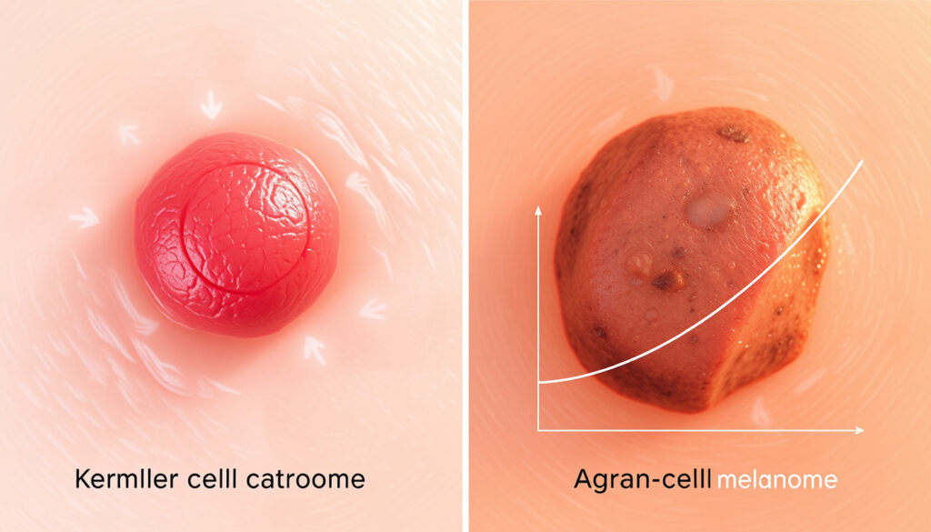
Merkel Cell Carcinoma vs Melanoma: Symptoms, Spread & Survival
If you’ve spotted a firm, fast-growing nodule on sun-exposed skin and found yourself down a rabbit hole of skin cancer types, you’re in the right place. Merkel cell carcinoma (MCC) is rare enough that most people have never heard of it—and aggressive enough that knowing what to look for actually matters. This guide cuts through the confusion, comparing MCC head-to-head with melanoma on the symptoms, spread patterns, and survival numbers that count most.
Annual US cases: about 3,000 · Common sites: head, neck, sun-exposed skin · Recurrence risk: high · Metastasis risk: often to lymph nodes · Associated virus: Merkel cell polyomavirus
Quick snapshot
- Rare neuroendocrine skin cancer (MD Anderson Cancer Center)
- Median patient age 75.5 years vs 57 for melanoma (PMC Comparative Study)
- Head and neck affected in 47% of cases vs 25.8% for melanoma (PMC Comparative Study)
- Exact percentage of cases directly caused by polyomavirus infection
- Long-term immunotherapy durability beyond 5-year data
- Incidence rose 0.8 per 100,000 in 2000–2021 SEER data (PMC 2024 SEER Analysis)
- Survival improved after checkpoint inhibitor approval in 2011 (PMC 2024 SEER Analysis)
- Immunotherapy expanding to earlier disease stages (Yale Medicine)
- Ongoing trials combining checkpoint inhibitors with targeted therapy (Yale Medicine)
| Attribute | Value |
|---|---|
| Cancer type | Neuroendocrine skin cancer |
| Incidence | 3,000 US cases yearly |
| Primary sites | Head/neck (50%) |
| 5-year survival | Stage-dependent: 75% localized |
| Median age at diagnosis | 75.5 years |
| Male predominance | 63% of cases |
What are the first signs of Merkel cell cancer?
Merkel cell carcinoma typically announces itself as a painless, firm nodule on sun-exposed skin. Most lesions appear on the head and neck, though arms and legs are also common locations. The nodule is often red or violet and may grow noticeably over just two to three months.
Initial appearance
The classic presentation is a rapidly enlarging, dome-shaped bump that rarely causes pain. About 88% of patients report the lesion was asymptomatic at first, meaning it drew attention purely from its appearance or unexpected growth rather than discomfort. The color ranges from red to purple, sometimes mimicking a blood blister or an innocent-looking pimple that won’t go away.
Growth characteristics
MCC tumors demonstrate rapid doubling times. Research from Frontiers in Oncology (tumor biology journal) documents growth over two to three months, with metastasis propensity reaching up to 35% of cases. This speed sets MCC apart from most non-melanoma skin cancers.
Associated symptoms
Beyond the visible nodule, some patients experience bleeding or ulceration as the tumor advances. The AEIOU mnemonic—Asymptomatic, Expanding rapidly, Immune suppression, Older than 50, UV-exposed site—captures the key warning signs clinicians use when evaluating suspicious lesions.
Unlike melanoma, which patients often learn to spot via the ABCDE rule, MCC has no widely known self-screening equivalent. The combination of rapid growth plus sun-exposed location should trigger a dermatologist visit within weeks, not months.
How serious is Merkel cell skin cancer?
Merkel cell carcinoma carries a reputation as one of the deadliest forms of skin cancer. The numbers back this up: MCC kills roughly one in three patients, making it the second leading cause of skin cancer death after melanoma. The danger escalates dramatically once the disease spreads beyond the original site.
Aggressiveness factors
MCC spreads early and often. Regional lymph node involvement occurs at diagnosis in 11.0% of MCC cases compared to just 3.1% for melanoma, according to PMC comparative research (NCBI clinical database). Distant metastases appear in 7.2% of MCC versus 2.1% for melanoma at initial presentation.
Recurrence rates
MCC recurs in a significant portion of patients even after seemingly successful treatment. Follow-up screening every three to six months during the first two years post-treatment is standard protocol, per Yale Medicine (academic medical center). The high recurrence rate makes long-term surveillance essential.
Mortality risks
Stage-specific survival data from SEER registry and published by the American Cancer Society (cancer data registry) shows five-year relative survival of 79% for localized disease, 66% for regional spread, and 31% for distant metastasis. The combined five-year survival rate sits around 69%.
Patients over 70 face the steepest odds. The cancer-specific mortality hazard ratio for MCC is 2.33 compared to melanoma—meaning MCC patients are more than twice as likely to die from their cancer than melanoma patients, adjusted for other factors.
How quickly does Merkel cancer spread?
MCC earns its “aggressive” label through rapid local growth and early lymph node invasion. Unlike some skin cancers that remain localized for years, MCC frequently disseminates within months of appearance, making prompt diagnosis and treatment critical.
Local spread
Tumors can double in size within weeks. The rapid expansion characteristic helps distinguish MCC from benign skin lesions, but this same quality means delays in treatment directly impact outcomes. Clinical guidelines emphasize urgent biopsy confirmation once a suspicious lesion shows fast growth on sun-exposed skin.
Lymph node involvement
Pathologic nodal status serves as a key predictor of survival outcomes, according to PMC comparative research (NCBI clinical database). Sentinel lymph node biopsy is typically recommended for staging, as microscopic spread often precedes clinically detectable node involvement.
Distant metastasis
When MCC metastasizes, it commonly targets distant skin sites, lungs, liver, and brain. Five-year survival drops to approximately 14% for stage IV disease versus 29% for stage IV melanoma, based on 2000–2021 SEER data analyzed in PMC 2024 comparative study (NCBI clinical database).
The combination of immune suppression and older age creates a particularly high-risk subgroup. Patients with organ transplants or chronic lymphocytic leukemia face substantially elevated metastasis risk and worse outcomes.
Where is the most common site of Merkel cell carcinoma?
MCC demonstrates a striking predilection for the head and neck region, occurring there in roughly half of all cases. This concentration on sun-exposed areas reinforces ultraviolet exposure as a key driver, though the virus connection adds another layer to the disease’s biology.
Head and neck
The face and scalp together account for 36% of MCC lesions, compared to just 14% for melanoma. The head and neck excess ratio (observed-to-expected) reaches 5.15 for MCC versus 2.47 for melanoma, based on PMC 2024 SEER analysis (NCBI clinical database). This anatomical pattern likely reflects cumulative UV exposure combined with other localized factors.
Arms and legs
The extremities rank second to head and neck for MCC occurrence. Upper limbs show higher rates than lower limbs, again correlating with sun exposure patterns. Unlike melanoma’s trunk predominance, MCC rarely originates on the torso.
Other sites
Rare presentations can occur on buttocks, genitals, or non-sun-exposed skin. These atypical locations account for a small fraction of cases but present diagnostic challenges since the clinical picture doesn’t match the typical sun-exposed pattern.
The anatomical distribution reveals something important: UV exposure alone doesn’t explain MCC. The Merkel cell polyomavirus lurks in normal skin cells, and researchers believe viral factors interact with sun damage to trigger malignancy in susceptible individuals.
Which is worse, melanoma or Merkel cell carcinoma?
The honest answer requires breaking down the comparison across multiple dimensions. MCC kills faster and spreads earlier, but melanoma affects far more people and carries its own serious risks. Looking at the data together tells a more nuanced story than a simple “which is worse” ranking.
Survival rates
The survival gap is stark. Ten-year cumulative survival stands at 17.7% for MCC versus 61.3% for melanoma, per PMC comparative research (NCBI clinical database). One-year survival rates show 57.7% for MCC compared to 92.9% for melanoma—a difference that translates to thousands of lives lost to MCC annually despite far fewer diagnoses.
Treatment responses
Both cancers benefited from immunotherapy advances after 2011. Checkpoint inhibitors now drive improved outcomes for metastatic disease in MCC and melanoma alike, documented in Merkel Cell Organization (patient advocacy and research summary). Yale Medicine oncologists report durable responses in patients with metastatic MCC receiving contemporary immunotherapy regimens.
Aggressiveness comparison
MCC behaves more aggressively on local and regional fronts—it invades lymph nodes earlier, recurs more frequently, and delivers worse stage-for-stage survival. However, melanoma’s higher incidence (27.3 versus 0.8 per 100,000 based on 2000–2021 SEER data) means it claims more total lives. The MD Anderson Cancer Center (cancer research and treatment institution) notes MCC ranks as the second leading cause of skin cancer death after melanoma.
Five key metrics show the divergence between these two skin cancers. Both share UV exposure as a risk factor, but MCC skews toward older males with a distinct anatomical pattern, while melanoma affects a broader age range with more even gender distribution.
| Metric | Merkel Cell Carcinoma | Melanoma |
|---|---|---|
| Incidence (per 100,000, 2000–2021) | 0.8 | 27.3 |
| Cases documented (2000–2021) | 19,444 | 646,619 |
| Median age at diagnosis | 75.5 years | 57 years |
| Head/neck involvement | 47% | 25.8% |
| Regional lymph node at diagnosis | 11.0% | 3.1% |
| 5-year overall survival | 60% | 93% |
| Stage IV 5-year survival | 14% | 29% |
| Cancer-specific mortality HR | 2.33 (reference) | Lower |
The implication: MCC patients face more than double the cancer-specific mortality risk compared to melanoma patients, adjusted for age and other clinical factors.
“With contemporary immunotherapy, many patients with metastatic Merkel cell carcinoma are living for years, and some might have complete, durable responses.” — Harriet Kluger, MD, Yale Medicine oncologist
“Many clinicians assume MCC and melanoma behave similarly. However, MCC occurs in an older population, more frequently on the head and neck, in a higher percentage of men.” — Researchers, PMC Comparative Study (NCBI clinical database)
The comparative data reveal two distinct profiles: MCC targets older patients with a narrow anatomical footprint and aggressive biology, while melanoma casts a wider net across ages and body locations. The immunotherapy era has improved survival for both, but the underlying gap persists.
For anyone noticing a rapidly growing nodule on sun-exposed skin—especially on the head or neck of an older adult—the message is clear: don’t wait and see. MCC rewards early intervention and punishes delay. Dermatologists have the tools to diagnose and stage it; the patient’s job is simply to show up when the signs appear.
Related reading: Deep Vein Thrombosis Symptoms
aacrjournals.org, onlinelibrary.wiley.com, theplasticsfella.com, psdermgroup.com, cancer.gov
While Merkel cell carcinoma grows rapidly without much color change, visual guide to melanoma signs illustrates classic melanoma features like asymmetry for better differentiation.
Frequently asked questions
What causes Merkel cell carcinoma?
MCC arises from a combination of factors including UV exposure, older age, and Merkel cell polyomavirus infection. The virus is present in most MCC tumors, though researchers are still determining its exact role in malignancy. Immunosuppression significantly elevates risk, particularly in organ transplant recipients and patients with chronic lymphocytic leukemia.
What is Merkel cell carcinoma virus?
Merkel cell polyomavirus (MCPyV) is a common virus found on normal skin. Most people carry it without harm, but in rare cases involving additional risk factors like UV damage and immune suppression, the virus may contribute to MCC development. The virus was discovered in 2008 and remains an active area of research.
What is the prognosis for Merkel cell carcinoma?
Five-year survival ranges from 79% for localized disease to 31% for distant metastasis, per SEER data published by the American Cancer Society. Age over 70, lymph node involvement, and delayed diagnosis all worsen prognosis. Immunotherapy has improved outcomes for advanced disease since 2011.
Is Merkel cell carcinoma hereditary?
MCC is not considered hereditary in the traditional sense. No consistent germline mutations have been identified as major drivers. The main risk factors are environmental (UV exposure, viral infection, immunosuppression) rather than inherited genetic patterns.
How is Merkel cell carcinoma staged?
Staging follows standard TNM criteria: tumor size and depth (T), regional lymph node involvement (N), and distant metastasis (M). Sentinel lymph node biopsy is typically performed to assess nodal status. Stage I-II indicates localized disease, Stage III involves regional nodes, and Stage IV means distant spread.
What are treatments for advanced Merkel cell carcinoma?
Immunotherapy with checkpoint inhibitors (avelumab, pembrolizumab) represents first-line treatment for metastatic MCC. Clinical trials continue exploring combination approaches. Localized disease typically involves surgery plus radiation. Treatment decisions depend on stage, patient age, and immune status.
Can Merkel cell carcinoma be prevented?
Sun protection reduces risk for MCC as for all skin cancers. Regular skin exams are particularly important for older adults and those with immunosuppression. No specific prevention exists for Merkel cell polyomavirus infection, which is widespread in the general population.
More related posts
 Izak Rankine: AFL Career, Background, Height, and Speed
 Kelsea Ballerini Biography: Songs, Relationships, Net Worth
 Kareena Kapoor: Marriages, Children, Health & FAQs
 Colin Fassnidge: Cocaine Driving Case & Career Update
 Katie Leung Biography: Child, Net Worth, and Ethnicity
 Jos Verstappen: F1 Career, Net Worth & Relationship with Max
 Kelsea Ballerini: Biography, Relationships & Music Career
 8 Ball Pool Unblocked – Top Sites and Play Tips